TMBL is well equipped with cutting edge instruments for characterizing biological and synthetic materials at all scales. Some examples are highlighted here, in addition to others avaialble in Biopolis and the National University of Singapore.
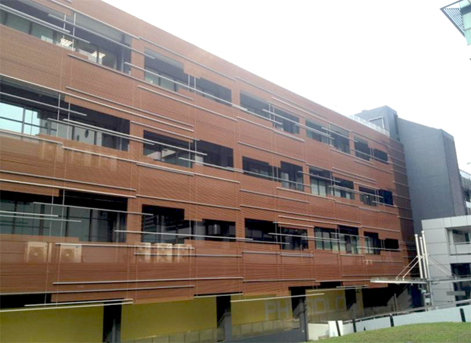
The location of the Confocal Microscopy Unit and Flow Cytometry Laboratory at the National University of Singapore (NUS).
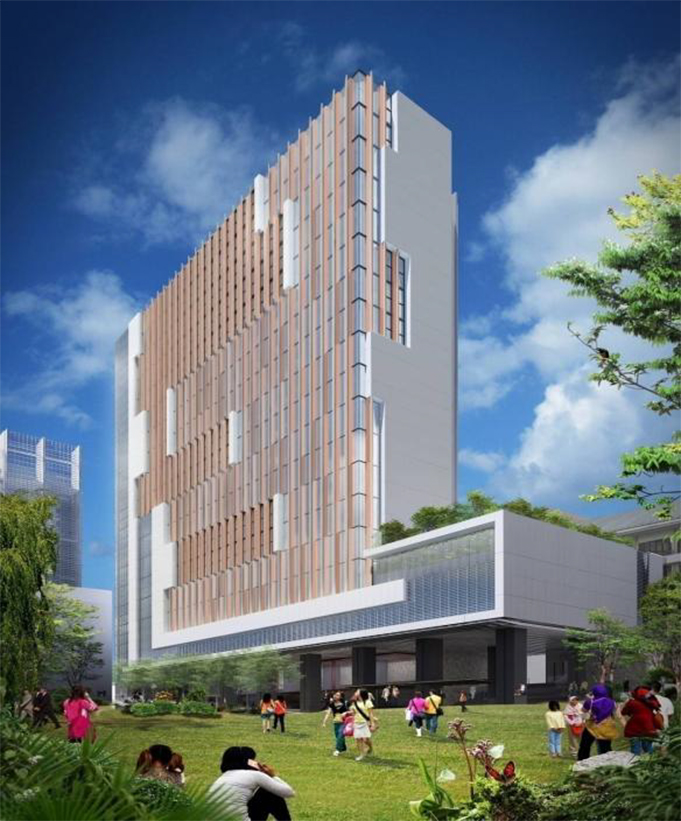
The location of the main research laboratory of Translational Mechanobiology Lab at the National University of Singapore (NUS).
Link to NUSMed Medical Science Cluster Research Core Facilities
Zeiss Axio Observer
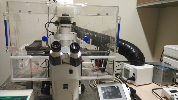
Gain accurate and reliable knowledge about living cells with your Axio Observer. According to the cell biology experiments you run, choose the degree of motorization and automation out of 3 different stands. Simply stay focused on your scientific experiments.
TissueGnostic: TissueFAXS Slide Scanner
The TissueGnostics TissueFAXS Slide Scanner can scan digital slides or images of tis sue sections, Tissue Microarrays (TMA), cell culture monolayers, smears and of other samples on slides and oversized slides, in Microtiter plates, Petri dishes and plates in both bright -field and epiflu orescence modes. TissueFAXS Laser equiped: 365nm, 488nm, 545nm, 643nm, 710nm.
Olympus FV3000 Confocal Microscope
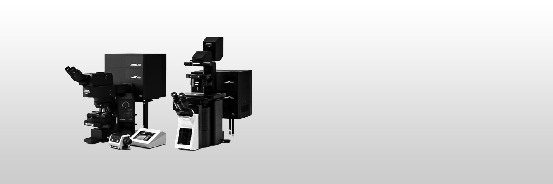
The Olympus FV3000 is an inverted IX83 laser scanning confocal microscope system equipped for specimens stained or labelled with up to 4 fluorophores of differing excitation wavelengths. It has all diode lasers and LED illumination. It has high sensitivity GaAsP Photomultiplier Tubes for high S/N ration images under very low excitation light. The system also has live spectral unmixing with TruSpectral detection. The system supports macro to micro imaging from 1.25X up to 100X, and users can employ image stitching to generate overview images. Being fitted with a resonant scanner which is ideal for high-speed imaging capturing at 30 frames per second, which also greatly reduces photobleaching and phototoxicity for live cell imaging. Accurate time-lapse imaging is ensured with Z-Drift Compensator keeping samples focus during an experiment despite changes in temperature or addition of reagents.
EVOS M7000 Brightfield Microscope
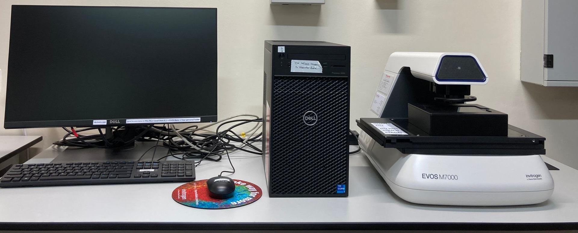
The EVOS M7000 microscope is a fully automated, no-personal-contact imaging system. It incorporates both monochrome and color high-resolution CMOS cameras for the best of both fluorescent and colorimetric imaging. The M7000 can scan multiwell plates automatically and features speedy autofocus, image acquisition, and large data processing.
Yokogawa CQ1 Spinning Disk Confocal Microscope
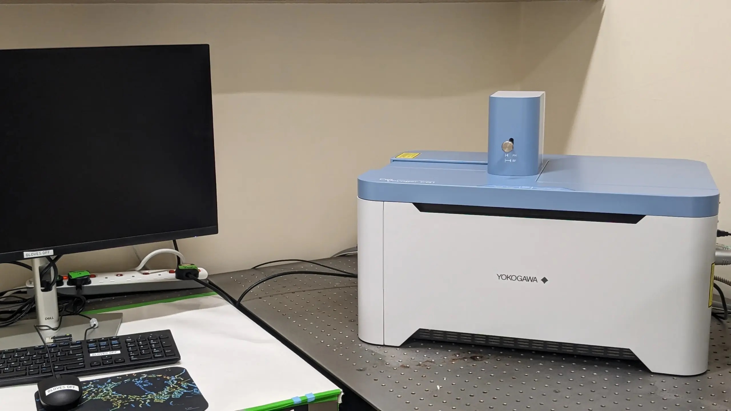
CQ1 Benchtop High-Content Analysis System enables 3D imaging and quantification of live cell clusters, such as spheroids within a 3D culture vessel, as they are, keeping the cells intact. CQ1 exports feature data in general formats which are readable by various third-party software for advanced data analysis. It is possible to construct fully customized CQ1-based system by integrating with external systems, via robot for culture dish handling.
Bitplane Imaris 8.1
Imaris’ three views - Arena, Surpass and Vantage - will naturally guide you through the stages of the Scientific Method. At your disposal you have a fully integrated platform which allows you to organize/explore your data, visualize it, (batch) analyze it, test hypotheses and present your conclusions in the best possible manner. Imaris 8.1 builds on the powerful and versatile infrastructure introduced with Imaris 8.0. The new version offers a range of useful solutions for microscopy core facilities and sites where users need to access and store their data using different workstations. - See more at: http://www.bitplane.com/imaris#sthash.dvsvKecy.dpuf
ImageJ

ImageJ is a public domain, Java-based image processing program developed at the National Institutes of Health.ImageJ was designed with an open architecture that provides extensibility via Java plugins and recordable macros. Custom acquisition, analysis and processing plugins can be developed using ImageJ's built-in editor and a Java compiler. User-written plugins make it possible to solve many image processing and analysis problems, from three-dimensional live-cell imaging to radiological image processing, multiple imaging system data comparisons to automated hematology systems. ImageJ's plugin architecture and built-in development environment has made it a popular platform for teaching image processing.
MATLAB
The MATLAB platform is optimized for solving engineering and scientific problems. The matrix-based MATLAB language is the world’s most natural way to express computational mathematics. Built-in graphics make it easy to visualize and gain insights from data. A vast library of prebuilt toolboxes lets you get started right away with algorithms essential to your domain. The desktop environment invites experimentation, exploration, and discovery. These MATLAB tools and capabilities are all rigorously tested and designed to work together.
Image Pro Plus
Image-Pro software includes the latest tools for scientific and industrial image analysis and image processing. Capture, process, measure, share, visualize and compare.
MetaMorph
MetaMorph® Microscopy Automation & Image Analysis Software is the industry standard for automated microscope acquisition, device control, and image analysis, bringing microscopists greater understanding of cell morphology, function, and behavior for over 25 years. It is the ideal "glue" for easily integrating dissimilar fluorescent microscope hardware and peripherals into a single custom workstation, while providing all the tools needed to perform meaningful analysis of acquired images. The software offers many user-friendly application modules for biology-specific analysis such as cell signaling, cell counting, and protein expression.
Link to NUSMed Medical Science Cluster Research Core Facilities
BD LSR Fortessa Flow Cytometry Analyser
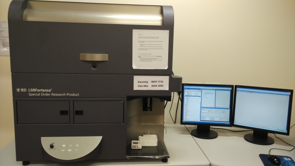
BD LSR Fortessa is equipped with four solid lasers (405nm, 488nm, 561m and 640nm). It can detect 16 colors simultaneously in addition to Forward Scatter (FSC) and Side Scatter (SSC). It can also perform 5-color analysis without compensation.
BC MoFlo Astrios EQ Sorter
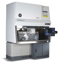
MoFlo Astrios EQ is able to perform 6-way sorting without compromise and measure crucial particle characteristics over more than three orders of magnitude, from as small as 200 nm to 30 µm diameter, all at the same time.
BC CytoFLEX
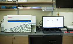
CytoFLEX LX B-R-V-Y-U-I expands research possibilities with up to six lasers and 21 color parameters. Six spatially separated lasers allows panels to be spread across the spectrum reducing cross talk and spectral overlap.
NanoFCM
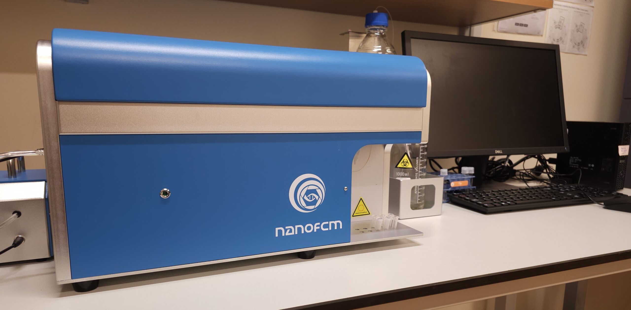
NanoFCM is equipped with two lasers (488nm and 640nm). It detects only nanoparticles below 1μm.
Agilent NovoCyte Advanteon
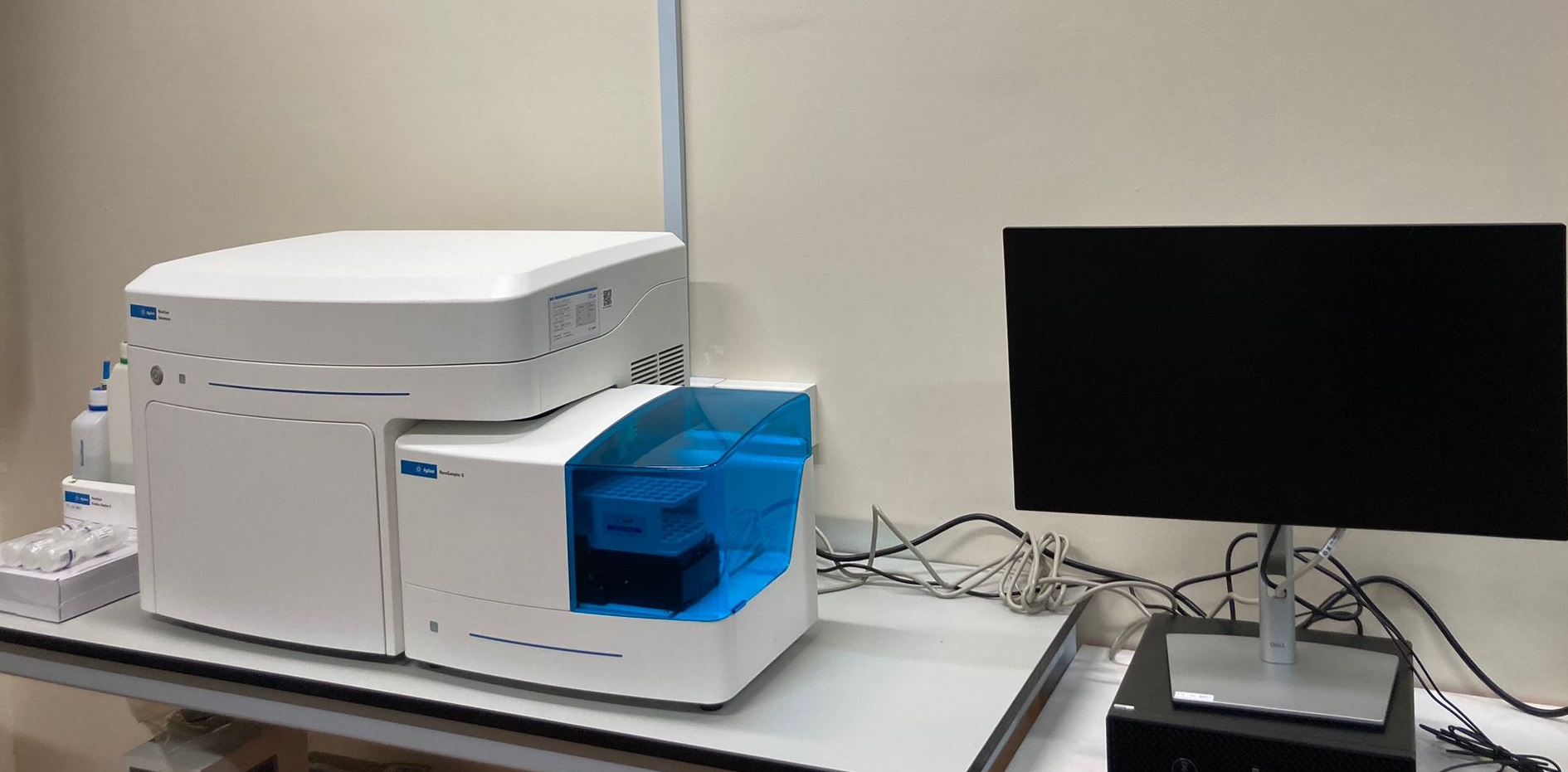
The NovoCyte Advanteon flow cytometer provides an advanced set of capabilities for the most demanding scientists, yet is remarkably simple to operate. The NovoCyte Advanteon can accommodate high-end and increasingly sophisticated multicolor flow cytometry assays. The system offers flexibility with 1, 2, or 3 laser options, up to 21 fluorescence channels, and 23 independent detectors. The sampler can efficiently process both FACS tubes (using a 40-tube rack) and 24-, 48-, 96-, and 384-well plates. For the NovoCyte Advanteon, the intuitive and industry honored NovoExpress software is now even more advanced, providing an exceptional user experience in data acquisition, analysis, and reporting.
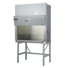
Biological Safety Cabinet Nuaire NU-425-300E
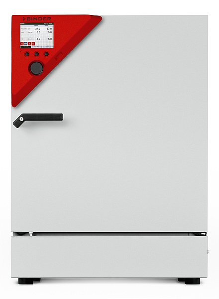
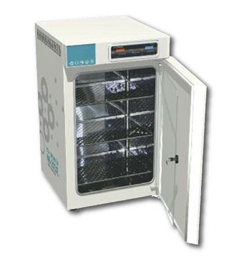
Cell Culture Incubator CB120
Roche LightCycler 1.5
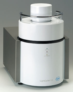
Roche LightCycler 1.5 provides all functionalities for running real-time PCR applications.
Tecan Infinite M1000
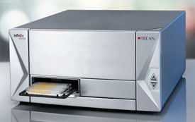
Tecan Infinite M1000 allows users to measure absorbance and fluorescence in a high-throughput manner.
Nanodrop ND2000
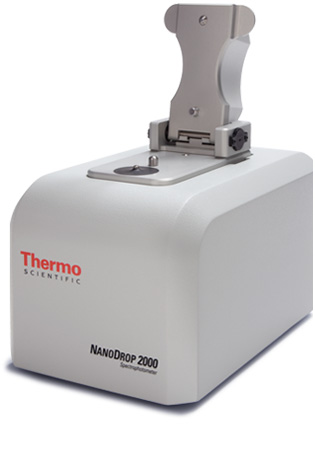
Nanodrop ND2000 measures DNA, RNA or protein concentration in less than 15 seconds.
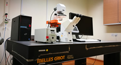
Olympus IX50 Phase Contrast Inverted Microscope
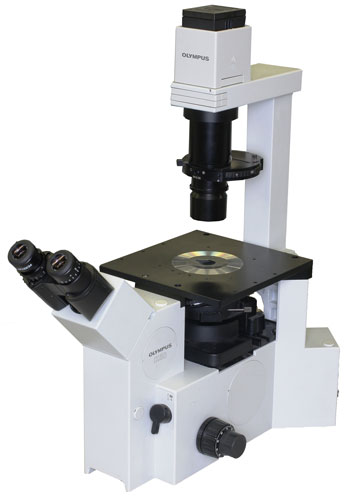
Olympus IX50 is a phase contrast inverted microscope that addresses the observation and imaging needs of high-level laboratory and clinical applications.
Bio-Rad T100 Thermal Cycler
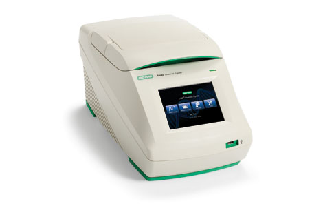
Bio-Rad T100 Thermal Cycler is a compact thermal cycler for running PCR.
Plasma Cleaner
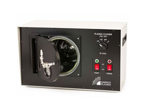
Harrick Plasma Expanded Plasma Cleaner for surface modification.
N-Biotek NB203QS
N-Biotek NB203QS Shaker CO2 Incubator for suspension cell culture.
Binder CB210
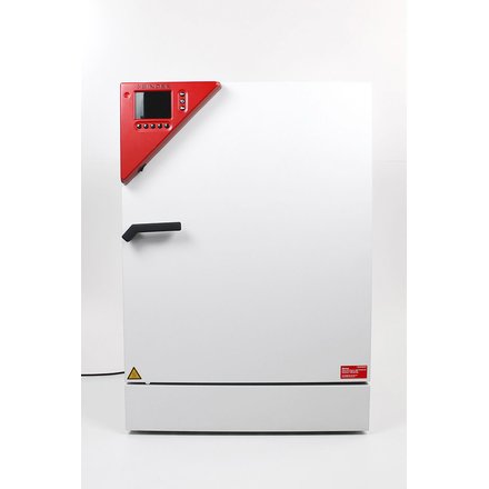
Binder CB210 Hypoxia Incubator for cell culture.